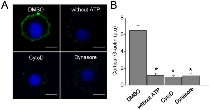
Figure 5. Inhibition of dynamin GTP-ase activity suppresses Ca2+-dependent de novo cortical actin polymerization.

A: Representative images of F-actin formation in cells permeabilized in the presence of 10 µM free Ca2+. Note that no new polymerized cortical actin was observed when the permeabilization was performed in the absence of ATP-Mg (n = 16) or in the presence of 4 µM CytoD (n = 27) or 100 µM dynasore (n = 28) Scale bar = 10 µm B: Quantification of G-actin staining mean intensity at the cell periphery. Data are means of cortical actin fluorescence intensity *p<0.05 compared with cells treated with DMSO (ANOVA).
