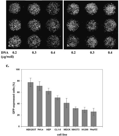
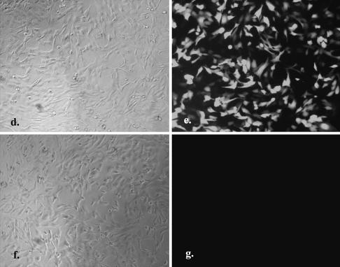
Figure 2.
Genes delivered into cultured cells in polymer-coated wells. The mini-wells (3 mm in diameter) were coated with a mixture of PEI (1 µg/cm2) and gelatin (1 µg/cm2). Cells (2000–8000) in culture medium containing 10% fetal calf serum and plasmid DNA were aliquoted directly into each well. The fluorescent image was taken 40 h after seeding the cells. The dose effect of pEGFP DNA (0.2, 0.3 and 0.4 µg/well, respectively) on the transfection of (a) HeLa and (b) 293T cells is shown in triplicate. Magnification 40×. (c) The relative transfection efficiency of various cell lines was quantified by counting GFP-expressed cells in the total cell population (mean ± standard deviation, obtained from triplicate wells). A similar experiment was performed in a 96-well plate coated with PEI/RGD-PEI/collagen (1.8, 0.18 and 0.3 µg/cm2, individually) mixture using HeLa cells (2 × 10⁴) and 1 µg of pEGFP (d and e) or pLuc (f and g). The fluorescent image was taken 24 h after seeding the cells. Magnification 100×. The experiments were repeated three times with similar results.


