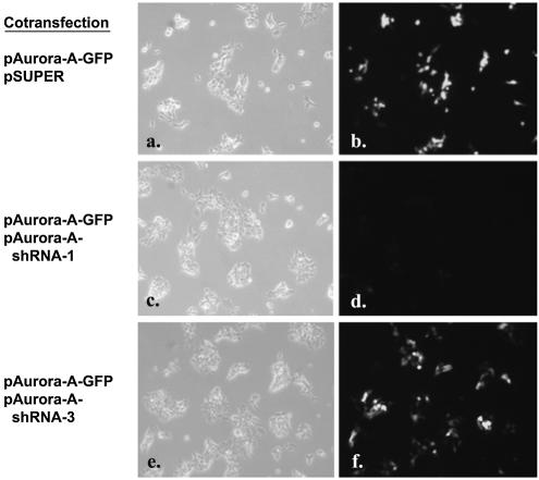
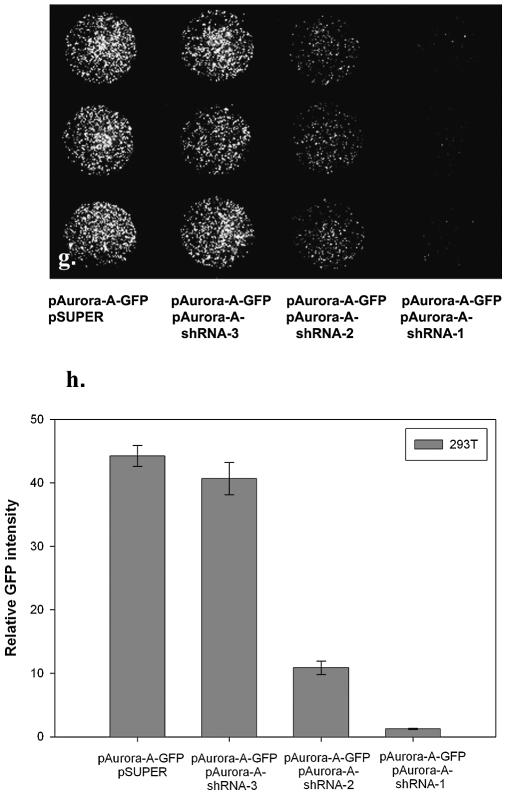
Figure 4.
shRNA-mediated gene silencing. Five thousand 293T cells in culture medium containing 10% fetal calf serum and plasmid DNA were aliquoted directly into the PEI/gelatin-coated wells (3 mm in diameter). These cells were cotransfected with equal amounts (0.15 µg of each plasmid per well) of reporter plasmid containing Aurora-A gene fused to GFP (pAurora-A-GFP) and pSUPER vector alone (a and b), pAurora-A-GFP and pAurora-A-shRNA-1 (c and d) or pAurora-A-GFP and pAurora-A-shRNA-3 (e and f). The fluorescent image was taken 40 h after seeding the cells. Magnification 100×. In a separate experiment, pAurora-A-GFP was cotransfected with pSUPER vector or with pAurora-A-shRNA-1, 2, 3, respectively. After transfection of 293T cells for 40 h, cells were subsequently fixed for 20 minutes in 3.7% paraformaldehyde solution. The fluorescent image (magnification 40×) was taken (g) and the relative fluorescent intensity was quantified using MetaMorph Imaging System software (h) (mean ± standard deviation, obtained from triplicate wells) with luciferase DNA (pLuc)-transfected cells as the background control. The experiment was repeated three times with similar results.


