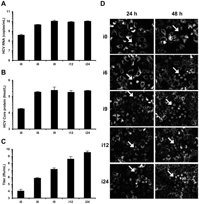
Figure 1. Increase of HCV titers after successive infections.
HuH-7 cells were electroporated in the presence of JFH1-CS-A4 RNA. Ten days later, the supernatant of electroporated cells was recovered (denoted supernatant i0) and used to perform successive infections in HuH-7-RFP-NLS-IPS. Each time the cells were 100% infected, the supernatant was recovered (supernatants recovered after “n” infection, denoted i1 to i24) and used to infect naive HuH-7-RFP-NLS-IPS cells. (A, B) The amount of HCV RNA (A) and Core protein (B) were quantified in these supernatants by RT-qPCR and fully automated chemiluminescent microparticle immunoassay, respectively. Results are expressed as HCV RNA copies/mL and fmol/L of HCV Core protein, respectively, and are reported as the mean ± S.D. of duplicate and triplicate measurements, respectively. (C) Viral titers were determined by ffu assay for i0, i6, i9, i12 and i24. Results are expressed as ffu/mL and are reported as the mean ± S.D. of three independent experiments. (D) HuH-7-RFP-NLS-IPS cells were inoculated with the different supernatants at low MOI. Foci of infected cells, identified by translocation of the cleavage product RFP-NLS to the nucleus, were visualized at 24 and 48 h. Images are representative of three independent experiments.

