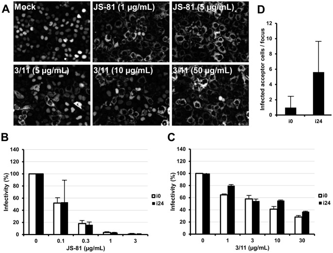
Figure 3. Viral entry of cell culture adapted HCV.
(A, B, C) Neutralization of cell culture adapted HCV with 3/11 and JS-81 MAbs. HuH-7-RFP-NLS-IPS cells were infected with i0 or i24 in the absence (Mock) or the presence of 3/11 anti-E2 or JS-81 anti-CD81 MAbs, at the indicated concentration. (A) Images taken 48 h after infection with i24 are representative of three independent experiments. (B, C) Intracellular HCV RNA was quantified 48 h after infection. Results are expressed as percentages of infectivity relative to infectivity in the absence of antibodies and are reported as the means ± S.D. of two independent experiments. (D) Cell-to-cell transmission of cell culture adapted HCV. Naive HuH-7-RFP-NLS-IPS cells (acceptor cells) were seeded with HuH-7-EGFP-IPS cells, infected with either i0 or i24 (donor cells). Cultures were treated with 50 µg/mL of the 3/11 anti-E2 neutralizing MAb to prevent cell-free infection. The results are expressed as the mean number of HCV infected acceptor cells/focus ± S.D., determined in 140 separate foci, 24 h post-seeding.

