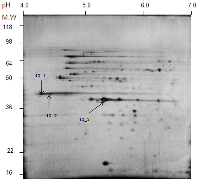
Figure 1. 2D-SDS-PAGE of S. flexneri 3a outer membrane proteins.
100 µg of crude preparation of the outer membrane proteins was precipitated with TCA. The pellet was washed with 12.5% TCA, acetone and air dried. The precipitate was dissolved in 125 µl of the sample buffer (pH 4–7). The IPG strip (11 cm) was soaked in the sample solution placed in the ceramic container provided with the IPGphor system. The first dimension of the electrophoretic separation was done according to the manufacturer’s recommendations. Proteins resolved on the buffer strip were reduced with DTT and alkylated with iodoacetamide. For the second dimension the IPG strip was placed over a vertical slab of 12.5% polyacrylamide gel prepared according to Laemmli [40]. Protein spots were visualized using the silver staining method as described by Shevchenko et al. [41]. Spots 13_1 and 13_2 were identified as OmpC and spot 13_3 as OmpA (Table 2).

