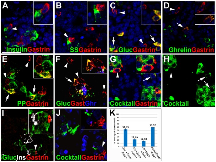
Figure 2. Presence of gastrin-expressing cells that do not stain for other islet hormones.
Images in panels A–H are from E15.5 mouse embryos. Arrows indicate cells co-expressing gastrin and other hormones, whereas arrowheads represent cells that express only gastrin. A–E. Co-staining for gastrin and insulin (A) or somatostatin (B) reveals no overlap. Co-staining for gastrin and glucagon (C), ghrelin (D) and pancreatic polypeptide (E) reveals some overlap, but some gastrin positive cells are negative for the other hormones. F. Triple staining for glucagon (green), gastrin (red) and ghrelin (blue) reveals some cells that stain only for gastrin. G–H. Co-staining for gastrin (red) and a cocktail of antibodies against insulin, glucagon, somatostatin, pancreatic polypeptide and ghrelin (G, H, green) reveals some cells that stain only for gastrin. I. Staining for gastrin (DAKO) reveals some gastrin+ cells that are negative for insulin and glucagon in e22 Psammomys obesus. J. Co-staining for gastrin (red) and a cocktail of antibodies against insulin, glucagon, somatostatin, pancreatic polypeptide and ghrelin (green) in e22 Psammomys obesus, reveals cells that stain only for gastrin. K. Quantification of cells expressing gastrin with glucagon, ghrelin, pancreatic polypeptide or all pancreatic hormenos as percentage of all cells expressing gastrin at E15.5 (at least 100 cells were counted from different pancreata (n>5)). Scale bar, 100 µm. Values are presented as mean ± SE.

