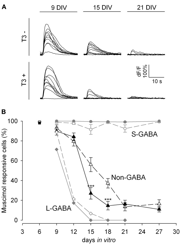
Figure 2.
GABA shift in T3+ and T3− networks shown by Fluo-4 recordings. (A). Representative calcium transients of non-GABA grown either in T3− and T3+ in response to muscimol application at 9, 15, and 21 DIV. (B). The developmental shift from depolarizing to hyperpolarizing actions of GABA is shown by the fraction of neurons with a significant increase in fluorescence after muscimol stimulation. At 6 DIV most neurons (black and white rectangles) show calcium transients after muscimol application. Only few L-GABA (diamonds) and non-GABA (triangles) can be activated by muscimol application after day 9, but all S-GABA (circles) respond to muscimol until day 27. GABAergic neuron response to muscimol was little affected by T3 treatment. In contrast, T3 treatment accelerates the developmental downregulation of depolarizing actions of muscimol in non-GABA (black triangles).

