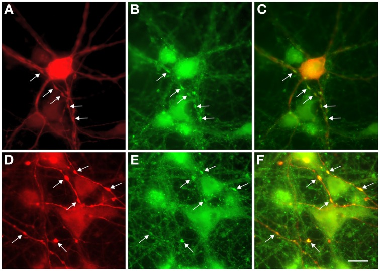
Figure 5.
Colocalization of GABA and synaptophysin immunostainings in 13-day-old cultures. (A,D) GABA staining shows prominent swellings along the axons of L-GABA neurons (arrows). (B,E). These boutons contain the synaptic protein synaptophysin. (C,F). Double exposure of (A,B) and (D,E) shows GABA/synaptophysin colocalization. Scale bar = 10 μm.

