
Fig. 4A–B.
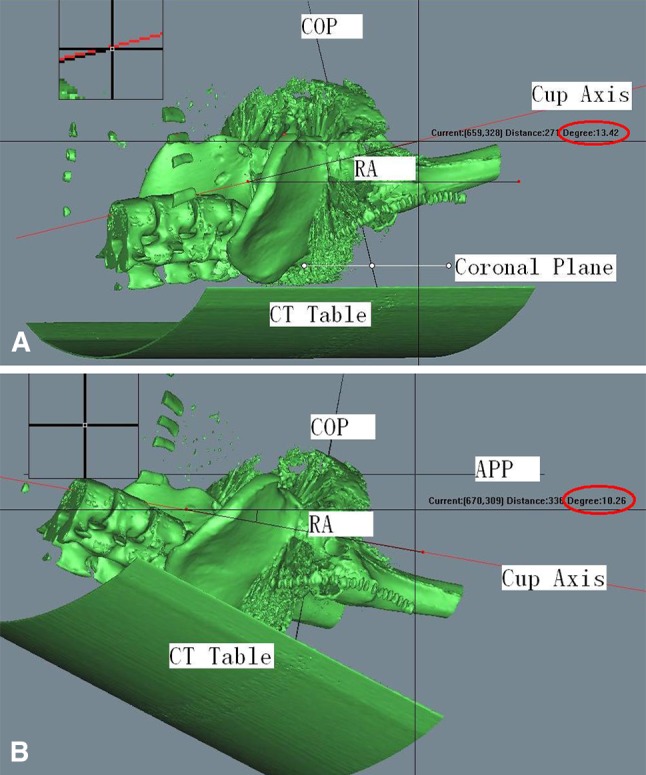
(A) Hip with ankylosing spondylitis and severe pelvic posterior tilt. Radiographic orientation, measured using the functional coronal plane, was in 13.4° of anteversion. (B) Hip with ankylosing spondylitis and severe pelvic posterior tilt. Radiographic orientation, measured using the APP as the coronal plane, was in 10.2° of retroversion. RA = radiographic anteversion; COP = cup opening plane.