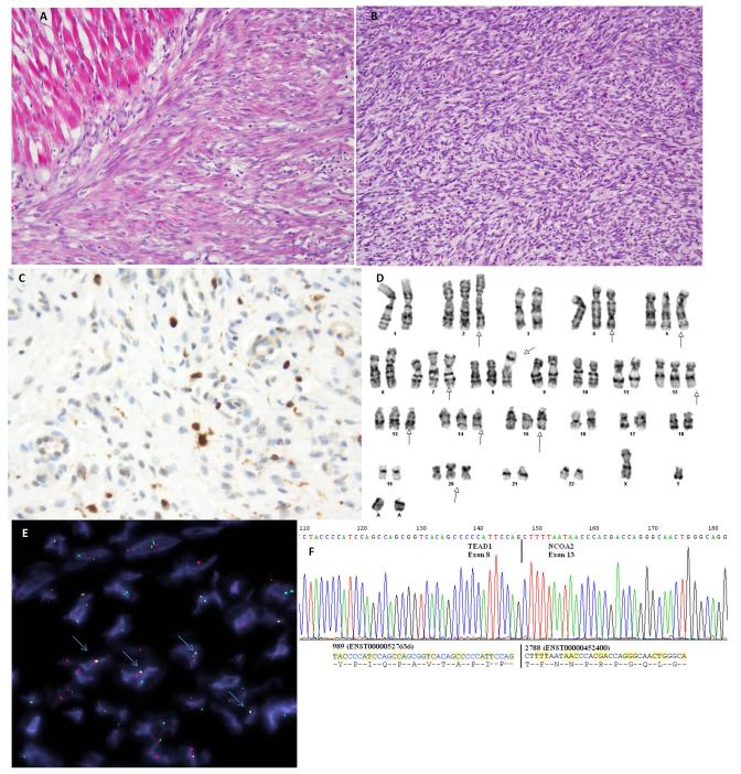
Figure 3. Spindle cell rhabdomyosarcoma with TEAD1-NCOA2 fusion.
Tumor from a 4 week-old baby boy who was born with a chest wall mass (case RMS4). (A) It is composed of short fascicles of spindle cells that have somewhat myofibroblastic appearance, infiltrating adjacent skeletal muscle. (B) A more cellular high grade component was also noted arranged in intersecting fascicles and associated with a high mitotic activity. Tumor cells demonstrate patchy positivity for Desmin and focal nuclear reactivity for myogenin (C) (A-C, original magnification 200×). (D) Partial karyotype showing a t(8;11) translocation. (E) Direct sequencing of the RT-PCR product confirmed the fusion of the TEAD1 exon 8 with NCOA2 exon 13. (F) FISH assay demonstrated TEAD1 and NCOA2 fusion-signal (highlighted with blue arrows; probes centromeric to NCOA2 in red, probes telomeric to TEAD1 in green).

