Figure 1.
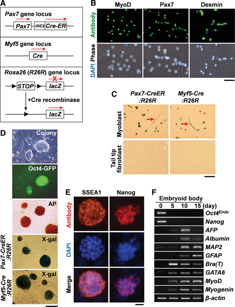
Generation of induced pluripotent stem cells (iPSCs) from primary myoblasts. (A): Schematic view of genetic lineage tracing strategy for Pax7-CreER:R26R and Myf5-Cre:R26R compound mice. (B): Panels show fluorescence views of myoblasts after antibody staining for MyoD, Pax7, and Desmin. Scale bar = 50 µm. (C): Culture of myoblasts from Pax7-CreER:R26R and Myf5-Cre:R26R compound mice shows lacZ+ permanently labeled cells including both myoblasts and multinucleated myotubes (arrows). By contrast, tail tip fibroblasts did not express lacZ. Scale bar = 100 µm. (D): ESC-like colonies were generated from myoblasts from Oct4-GFP mice. These colonies were positive for GFP and AP staining. Colonies generated from myoblasts isolated from Pax7-CreER:R26R and Myf5-Cre:R26R were positive for X-gal staining. Scale bar = 200 µm. (E): The myoblast-derived colonies expressed SSEA1 and Nanog. Scale bar = 20 µm. (F): Reverse transcription-polymerase chain reaction experiments show that embryonic bodies generated from myoblast-derived iPSCs expressed differentiation markers for three germ layers. AFP and Bra (T) denote α-fetoprotein and brachyury (T) genes, respectively. β-actin was monitored as a loading control. Nuclei were counterstained with DAPI (blue). Abbreviations: AP, alkaline phosphatase; DAPI, 4′,6-diamidino-2-phenylindole; GFP, green fluorescent protein; SSEA1, stage specific embryonic antigen-1. [Color figure can be viewed in the online issue, which is available at wileyonlinelibrary.com.]

