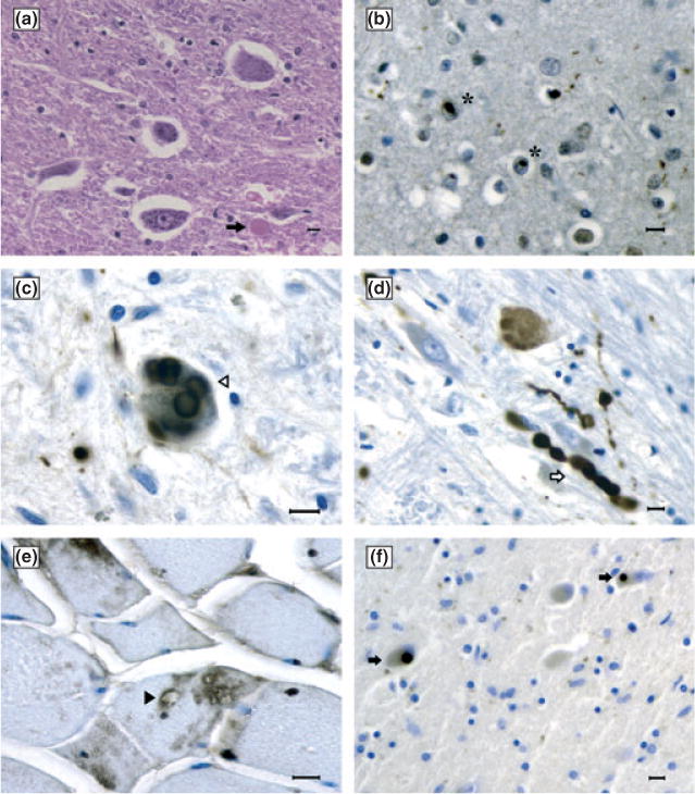
Figure 2.

(a) Neuronal loss and a rare axonal spheroid (arrow) in the anterior horn of the cervical cord (hematoxylin and eosin). (b) TDP-43-immunoreactive (IR) neuronal intranuclear inclusion in the frontal cortex (stars). (c) Alpha-synuclein-IR Lewy bodies (open arrowhead) and Lewy neuritis (open arrow) (d) in the substantia nigra. (e) Sarcoplasmic vacuoles (solid arrowhead) and VCP-IR deposits in the muscle. (f) TDP-43-IR Lewy-body-like inclusions in the thalamus (solid arrows). (a)–(e) Ohio family’s proband. (f) Pennsylvania family’s proband. Bars: 10 μm.
