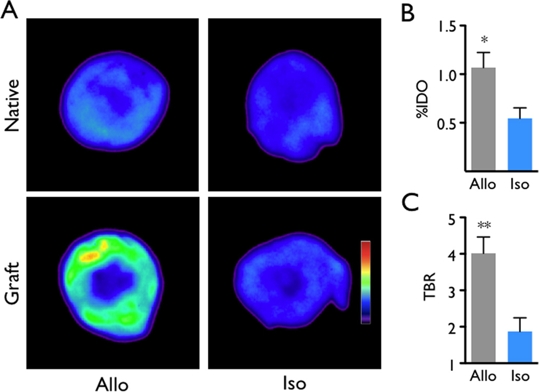
Figure 2.
Ex vivo validation of rejection imaging by autoradiography of native and transplanted hearts. (A) Representative autoradiographic images showed higher signals in allografts. (B) Scintillation counting of explanted grafts yields higher percent injected dose per organ (%IDO) in allografts. (C) Target to background ratio (TBR) of grafted hearts. The native heart served as background. Data are mean ± SEM, *p<0.05, **p<0.01.

