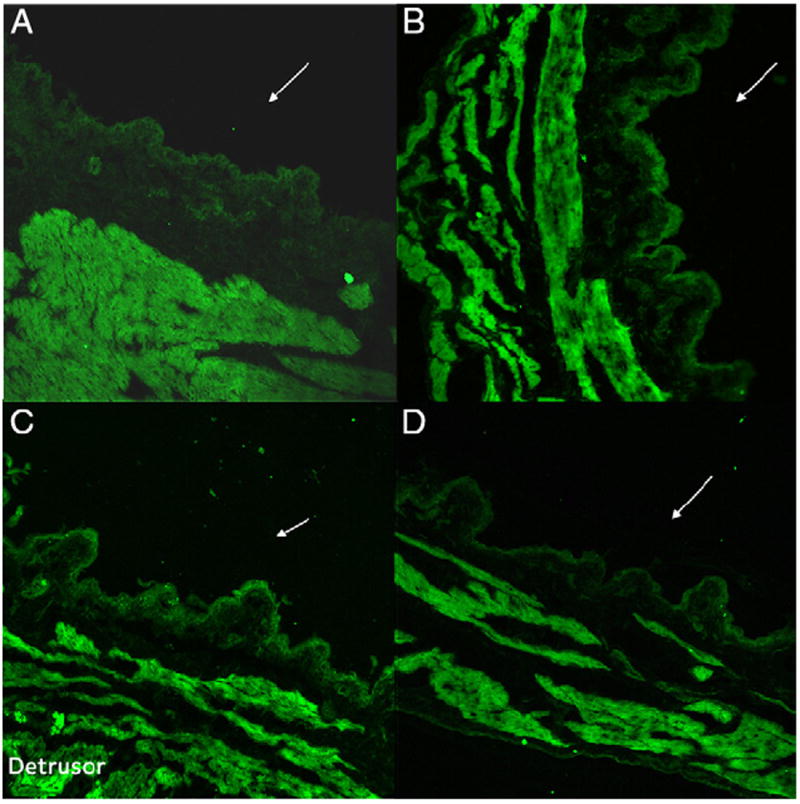
Figure 6.

Representative immunofluorescence labeling in rat bladder cross sections reveals that density of NGF immunoreactivity (green areas) was highest in detrusor in all groups. It increased after AA treatment in accordance with NGF expression in muscle. NGF immunoreactivity in bladder mucosa containing urothelium cell layer was absent in rats untreated with AA (A). It was noted in sham (C) and scrambled OND (B) treated rats. NGF immunoreactivity in urothelial cell layer was reduced in rats treated with antisense OND complexed with liposomes (D) to levels comparable to controls (A). Reduced from ×20.
