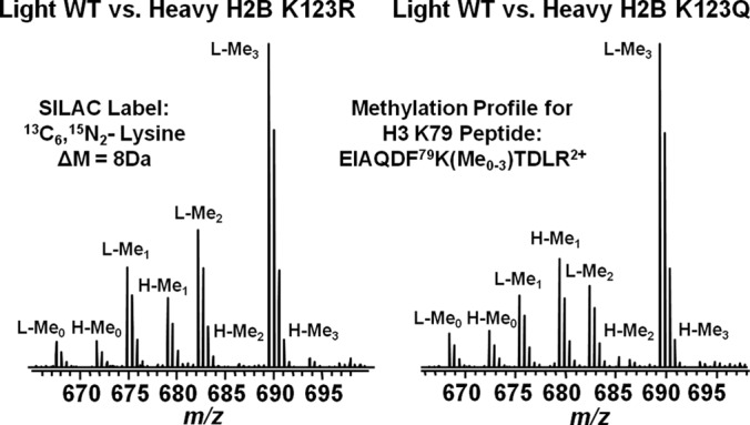
Fig. 2.

Mass spectrometry methylation profiles for the H3 K79 peptide fragment EIAQDF79K(Me0–3)TDLR2+ from SILAC light wild type:heavy mutant H2B K123R (left) and light wild type:heavy mutant H2B K123Q (right). Light and heavy peaks are labeled with L and H respectively. Peaks representing the unmethylated, mono-methylated, di-methylated or tri-methylated form of H3 K79 peptides are annotated by Me0, Me1, Me2, or Me3.
