
Fig. 2.
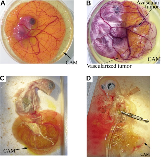
Biotinylation in vivo in the live chick. The chick embryo was grown in a Petri dish until E16 was perfused with Ringer's solution to eliminate blood cells and plasma proteins, which allowed subsequent biotinylation. A, chicken embryo and extra-embryonic membranes at day 6 of development. B, implantated tumor (U87) on the CAM on day 16 of development. C, pulling the embryo inside-out through the CAM opening submerged in the buffer bath allowed direct access to chicken chest with minimal injury to the tissue. D, cannulation of the heart and perfusion.