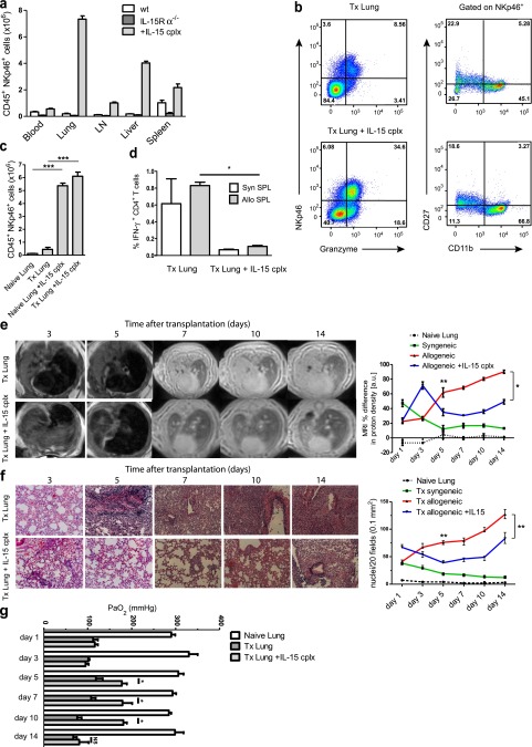
Figure 2.
Natural killer (NK)-cell expansion by IL-15 complexes favors transplantation outcome. (a) Amount of CD45+NKp46+ cell content in the indicated tissues from untreated, IL-15Rα(IL-15 receptor, α chain)−/−, and IL-15 complex–treated wild-type C57BL/6 mice (lung cells from three mice each are shown, means ± SEM; LN = cervical and mediastinal lymph nodes). (b) Flow cytometric analysis of CD45+NKp46+ lung cells from transplanted mice with and without IL-15 complex treatment stained for granzyme B, CD27, and CD11b expression by NKp46+ NK cells (representative data of six mice per group). (c) Lung cells gated on CD45+NKp46+ cells from naive and transplanted mice compared with IL-15 complex–treated mice (Tx Lung: Day 3 after transplantation, six mice per group, means ± SEM, ***P < 0.001). (d) Total numbers of IFN-γ-producing CD4+ T cells from transplanted lungs with (Tx IL-15 complex [cplx] lung) and without (Tx lung) IL-15 complex treatment of the recipient after 3 days of culture with syngeneic (syn SPL) or allogeneic (allo SPL) T cell-depleted splenocytes (composite data of three independent experiments, three mice per group, means ± SEM, *P < 0.05). (e) Representative magnetic resonance images from allotransplanted and IL-15 complex–treated allotransplanted mice on Days 3, 5, 7, 10, and 14 after transplantation. Infiltrations into lungs were assessed in four regions of interest for each lung and expressed by proton density in arbitrary units comparing transplanted with contralateral, normal lungs (percent differences). Naive lungs and syngeneically transplanted mice were also assessed (seven mice per group for Days 3 and 5, three mice per group for Days 7, 10, and 14 are shown, means ± SEM, *P < 0.05, **P < 0.01). (f) Representative lung sections stained with hematoxylin and eosin in untreated and IL-15 complex–stimulated allotransplanted mice on Days 3, 5, 7, 10, and 14 after transplantation. Quantification of the histologic analysis was performed by counting nuclei of infiltrating cells in 20 fields, each measuring 0.1 mm2 (six independent experiments per group for Days 3 and 5, three independent experiments per group for Days 7, 10, and 14 are shown, means ± SEM, Mann-Whitney, **P < 0.01). (g) Oxygenation measurements from blood samples taken from the left pulmonary vein at the indicated time points after Tx are shown for naive (naive lung), transplanted (Tx lung), and IL-15 complex–treated recipient of transplanted (Tx lung + IL-15 cplx) lungs (three independent experiments per group are shown, means ± SEM, Mann-Whitney, *P < 0.05, NS = no statistical significance).

