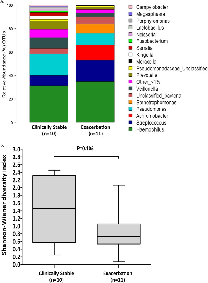
Figure 3.
(a) Comparison of the percent abundance of the major identified phyla in pooled samples collected from patients when clinically stable (cross-sectional study, n = 10) and at the start of treatment for an exacerbation (longitudinal study, n = 11). Similar patterns of phyla distribution were observed in both groups. OTUs = operational taxonomic units. (b) Box plot comparison of microbial diversity (Shannon-Wiener diversity index) in samples from patients when clinically stable (cross-sectional study, n = 10) and at the start of treatment for an exacerbation (longitudinal study, n = 11), where higher values correspond to higher diversity. The top and bottom boundaries of each box indicate 75th and 25th quartile values, respectively, with the black line inside each box representing the median (50th quartile). The ends of the whiskers indicate the range. No significant difference (P = 0.105, Mann-Whitney test) in microbial community diversity is apparent between the two groups.

