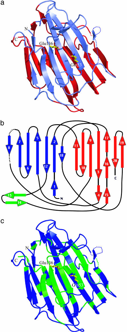
Fig. 2.
(a) Ribbon representation of SCP-B. The secondary structure assignment was determined by Database of Secondary Structure of Proteins (31). This drawing and Figs. 2c, 3, 4, 5, and 6b were produced by the program pymol (32). The active-site residues, Gln-53 and Glu-136, are shown in stick representation. The upper sheet is slate, and the lower sheet is red. (b) A topological diagram representing the secondary structure in SCP-B. The upper sheet harboring the active site is blue, and the lower sheet is red. The loop unique to SCP-B that is adjacent to the active site is shown in green. Seven loops cross from the upper to the lower sheet or from the lower to the upper sheet in this topography. (c) A representation of the regions of SCP-B that are highly conserved among the six members of the eqolisin family. Those segments of the chain corresponding to these highly conserved regions of SCP-B (asterisks in Fig. 1) are green.

