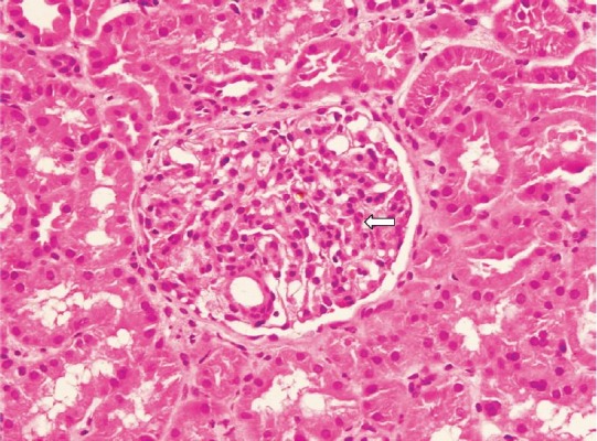
Fig. 1.

Light microscopic photomicrograph of kidney biopsy sample of a patient with renal involvement showing glomeruli with mesangial cell proliferations compatible with mesangioproliferative glomerulonephritis (H & E stain, 400X). Arrow shows glomeruli.
