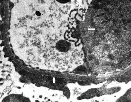
Fig. 3.

Electron microscopic photograph of kidney biopsy sample of patient with renal involvement showing endothelial cell swelling left point (white arrow) with degenerative changes and it also shows effacement of foot process upward pointing (white arrow) indicative of the podocyte injury/podocytopathy (40,000X).
