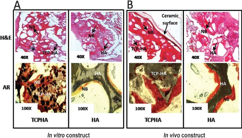
Fig. 2.

Light microscopic images of HA-SMC and TCP/HA-SMC three dimensional construct implanted in mice. (A) TCP/HA-SMC construct revealed new bone (NB) formation with some ceramics and fibrous tissue still seen. More bone area detected as compared to HA-SMC in both staining, H&E and Alizarin Red. (B) formation of new bone with a high surface area, and cell-shaped void embedded in the bone for TCP/HA-SMC construct. In HA-SMC construct there were lots of fibrous intrusion. AR staining confirmed more mineral activity in in vivo level both HA-SMC and TCP/HA-SMC Constructs.
