Fig. 1.

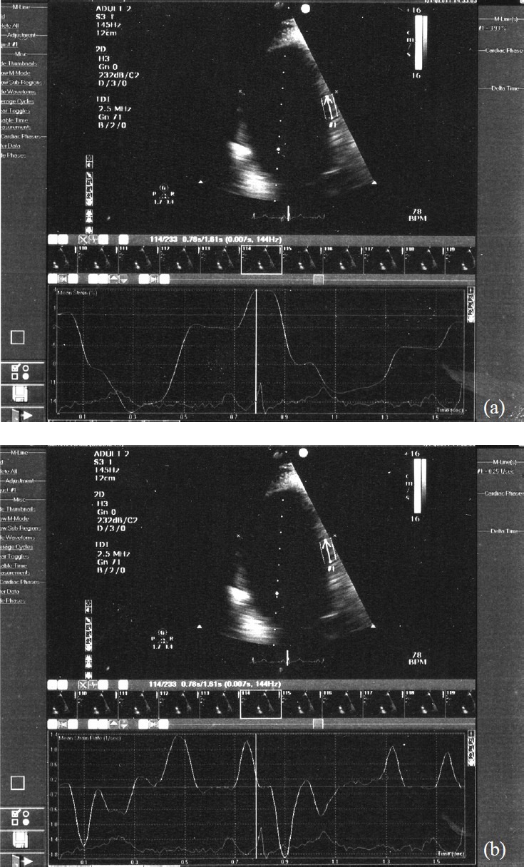
Four chamber view images with strain (a) and strain rate (b) analysis of the mid segment of interventricular septum in a control subject.

Four chamber view images with strain (a) and strain rate (b) analysis of the mid segment of interventricular septum in a control subject.