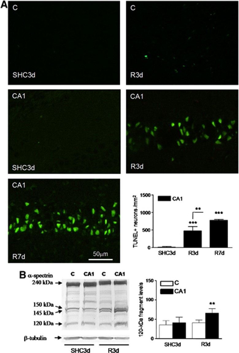
Figure 1.
Nuclear labeling for apoptosis detection in the hippocampal cornu ammonis 1 (CA1) region. (A) Brain sections of the cerebral cortex (C) or hippocampal CA1 region from control (SHC3d) and ischemic animals with reperfusion for 3 or 7 days (R3d and R7d, respectively) were used for apoptosis detection by the transferase-mediated dUTP nick-end labeling (TUNEL) assay. The figures are representative results. TUNEL-positive neurons were counted in CA1 from three different animals; error bars indicate s.d. (bar graph). ***P⩽0.001, compared with SHC3d control; **P⩽0.01, R3d compared with R7d; analysis of variance (ANOVA), P⩽0.0001. (B) Caspase-3 activation in the cerebral cortex and hippocampal CA1 region from ischemic animals. Samples of C or CA1 from SHC3d and R3d were subjected to sodium dodecyl sulfate-polyacrylamide gel electrophoresis and western blotting with anti-α-spectrin antibody. Arrows indicate the relative position for α-spectrin and their 150-, 145- and 120-kDa fragments. Data are the quantification of caspase-3-dependent 120-kDa fragment (bar graph) from three to six different animals; error bars indicate s.d. **P⩽0.01, compared with CA1 R3d; ANOVA, P=0.001.

