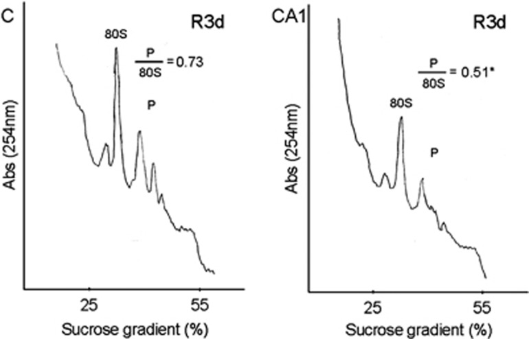
Figure 3.
Polysome dissociation upon reperfusion in the hippocampal cornu ammonis 1 (CA1) region. Polysome profiles were obtained from samples of the cerebral cortex (C) or hippocampal CA1 region (CA1) from ischemic animals with 3-day reperfusion (R3d). The values showed are the ratio of polysome (P)/80S species. The figures are representative results of three independent experiments. *P⩽0.05, CA1 compared with C.

