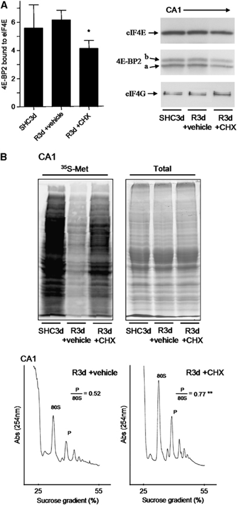
Figure 6.
Cycloheximide (CHX) prevents the binding of 4E-BP2 to eIF4E and restores protein synthesis in the hippocampal cornu ammonis 1 (CA1) region. (A) Binding assay of 4E-BP2 to eIF4E. Samples of the hippocampal CA1 region from controls (SHC3d) and untreated (vehicle) or treated animals with 1 mg/kg CHX that underwent ischemia with 3-dayreperfusion (R3d +vehicle and R3d +CHX, respectively) were loaded into m7GTP-Sepharose and subjected to sodium dodecyl sulfate-polyacrylamide gel electrophoresis (SDS-PAGE) followed by western blotting for anti-eIF4E (eIF4E), anti-4E-BP2 (4E-BP2), and anti-eIF4G (eIF4G) antibodies. The figure is a representative result from three different animals and shows the 4E-BP2 bound to eIF4E in the different experimental conditions. Arrows show the relative position for eIF4E, 4E-BP2 forms, and eIF4G. Data are the quantification of 4E-BP2 (a+b forms) with respect to eIF4E levels (ratios) (bar graph). Error bars indicate s.d. *P⩽0.05 compared with R3d +vehicle; analysis of variance, P⩽0.05. (B) Evaluation of protein synthesis rates. Hippocampal slices from SHC3d, R3d +vehicle, and R3d +CHX animals were incubated with [35S]Met for protein synthesis labeling and after the CA1s were processed. The extracts were resolved by SDS-PAGE, transferred onto nitrocellulose membranes and then exposed on phosphoimager to obtain an autoradiography of [35S]Met incorporated into proteins (upper-left image). Twin gels stained with Coomassie Blue were performed to the quantification of total protein (upper-right image). Polysome profiles were obtained from samples of R3d +vehicle (lower-left image) and R3d +CHX animals (lower-right image). The values are the ratio of polysome (P)/80S species. **P⩽0.01, R3d +CHX compared with R3d +vehicle. The figures are representative results from three different animals.

