Figure 1.
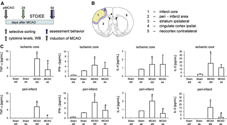
Enriched environment (EE) suppressed the inflammatory response after experimental stroke. (A) Experimental design. Spontaneous hypertensive rats were subjected to permanent occlusion of the middle cerebral artery (pMCAO). Two days later, neurologic deficit was evaluated by the rotating pole test and only animals with substantial deficits (test score 2 or lower) were randomized either into housing in standard (STD) laboratory cages or EE for 3 consecutive days. At day 5, after pMCAO, neurologic score was evaluated and brains were analyzed for proinflammatory cytokines. (B) Brain areas analyzed for proinflammatory cytokines: (1) infarct core; (2) peri-infarct area; (3) striatum-injured hemisphere; (4) cingulate cortex-injured hemisphere; (5) neocortex-noninjured hemisphere; (C) Levels of the proinflammatory cytokines TNF-α, IFN-γ, IL-4, and IL-5 in the infarct core and the adjacent peri-infarct area of rats housed either in STD (n=8) or EE (n=8) for 3 consecutive days after pMCAO and respective sham-operated rats (sham STD, n=6; sham EE, n=6). Data are presented as means±standard deviation (s.d.). The asterisk denotes statistical difference compared with all other experimental groups (*P<0.05), and the hash denotes statistical difference (#P<0.05) to the MCAO STD group (one-way analysis of variance, Bonferroni correction, Fisher's least significant difference test for levels of IL-5 in the MCAO STD group compared with the sham EE group). Please note that levels of cytokines measured in brain regions 3 to 5 are displayed in Supplementary Figure 1.

