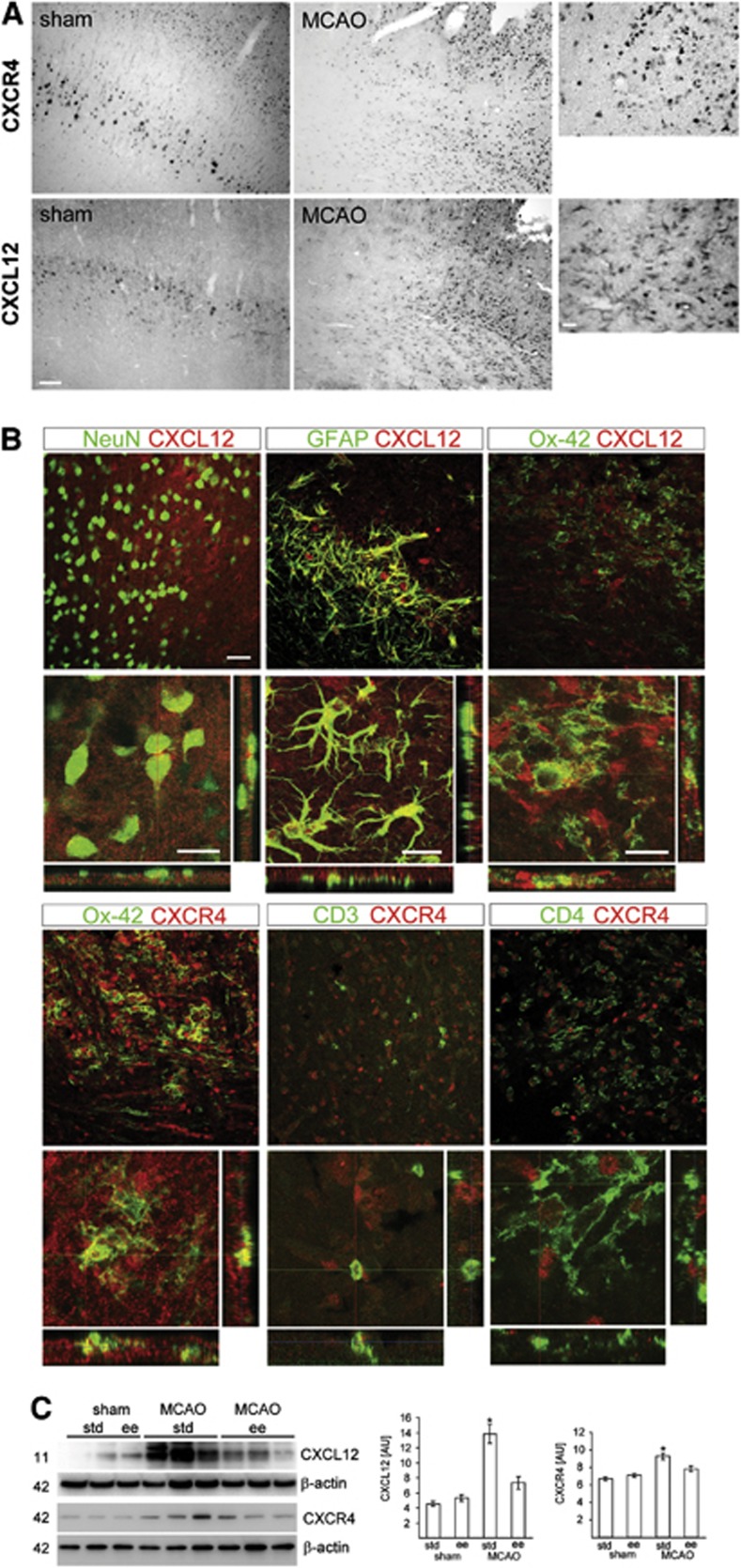
Figure 2.
Chemokine receptor CXCL12 pathway was suppressed by enriched environment (EE) after permanent middle cerebral artery occlusion (pMCAO). (A) Expression of CXCL12 and CXCR4 in the neocortex of sham-operated rats and the peri-infarct area of the ischemic hemisphere after pMCAO. Scale bars, low magnification, 100 μm; high magnification, 20 μm. (B) Identification of CXCL12- and CXCR4-expressing cells in the ischemic hemisphere. CXCL12 (Cy3, red) is expressed in NeuN+ (Cy5, green) neurons, GFAP+ (Cy5, green) astrocytes, and Ox-42+ (Cy5, green) microglia/macrophages. Colocalization of CXCR4 (Cy3, red) with Ox-42 (Cy5, green), CD3 (Cy5, green), CD4 (Cy5, green). Scale bars, 50 μm. (C) Levels for CXCL12 and CXCR4 at the infarct border zone from rats housed in standard (STD) (n=8) or EE (n=8) for 3 days after pMCAO or sham operation (sham STD, n=5; sham EE, n=5). Values are presented as means±s.d. and were calculated as percentage of β-actin expression. *P<0.05 versus all other experimental groups. One-way analysis of variance, Bonferroni correction.

