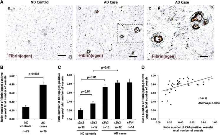
Figure 1.
Increased cerebrovascular deposition of fibrin(ogen) in Alzheimer's disease (AD) brains. Immunohistochemical staining of fibrin(ogen) was performed on cortical brain sections, and a ratio of the number of fibrin(ogen)-positive vessels versus the total number of vessels quantified was determined. (A) Representative images of fibrin(ogen) staining in non-demented (ND) (a) and AD (b) brains. Scale bar, 100 μm. (c) Higher magnification of fibrin(ogen)-positive vessels marked with dashed line in (b). Scale bar, 20 μm. (B) AD cases presented with significantly more fibrin(ogen)-positive vessels compared with ND controls (P=0.008). (C) In ND brains, apolipoprotein E (APOE) ɛ3/ɛ3 cases presented with significantly more fibrin(ogen)-positive vessels compared with APOE ɛ2/ɛ3 cases (P=0.04). In AD brains, no significant difference was observed in the number of fibrin(ogen)-positive vessels between the different APOE genotypes. Differences between groups were evaluated by unpaired Student's t-test (B) or one-way analysis of variance (ANOVA) and Tukey's post-hoc test (C). Values are presented as mean±s.e.m. (D) Simple linear regression analysis revealed a significant correlation between the number of fibrin(ogen)-positive vessels and the number of cerebral amyloid angiopathy (CAA)-positive vessels in the frontal cortex of AD cases (r2=0.31, ANOVA P=0.0004).

