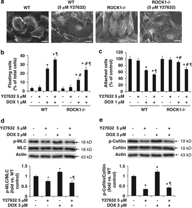
Figure 5.
Disruption of stress fibers by ROCK pan-inhibitor facilitates doxorubicin-induced detachment and death of attached cells. (a) Representative images of rhodamine-phalloidin staining for F-actin of WT and ROCK1-deficient cells treated with 5 μM Y27632 for 4 h. Bar, 50 μm. (b and c) Floating cells and attached cells were collected and counted after treatment with 1 μM doxorubicin and/or 5 μM Y27632 for 16 h. (d and e) Representative image (top) and quantitative analysis (bottom) of western blot of p-MLC2 and MLC2, or p-cofilin and cofilin in cell lysates from attached WT MEFs treated with 3 μM doxorubicin and/or 5 μM Y27632 for 30 min. The ratio of p-MLC to MLC or p-cofilin to cofilin was expressed as fold change relative to WT control. *P<0.05 versus control of the same genotype. #P<0.05 versus WT under the same treatment condition. ¶P<0.05 versus WT under doxorubicin only condition

