Figure 7.
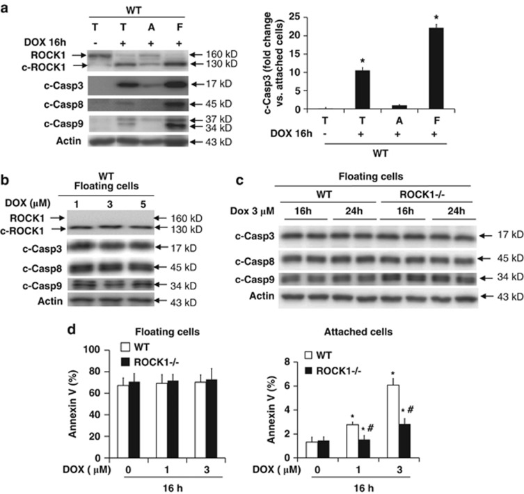
ROCK1 deletion does not inhibit apoptosis in detached cells. (a) Representative image (left panel) of western blot of ROCK1, cleaved caspase-3, -8, and -9 in cell lysates from total (T), attached (A) and floating (F) WT cells treated for 16 h with 3 μM doxorubicin. Densitometry analysis (right panel) of immunoreactive bands of cleaved caspase-3. Expression of cleaved caspase-3 was expressed as fold change relative to attached cells after doxorubicin treatment. *P<0.05 versus doxorubicin-treated attached cells. (b) Representative image of western blot of cleaved caspase-3, -8, and -9 in cell lysates from floating WT cells collected after 16 h of treatment with increasing dosages of doxorubicin as indicated. (c) Representative image of western blot of cleaved caspase-3, -8, and -9 in cell lysates from floating WT and ROCK1−/− MEFs collected after 16 h or 24 h treatment with 3 μM doxorubicin. (d) Apoptotic cell ratio was determined by annexin V staining followed by FACS analysis in floating (left) and attached (right) cells collected after treatment for 16 h with increasing dosages of doxorubicin as indicated. *P<0.05 versus control of the same genotype. #P<0.05 versus WT under the same treatment condition

