Figure 1.
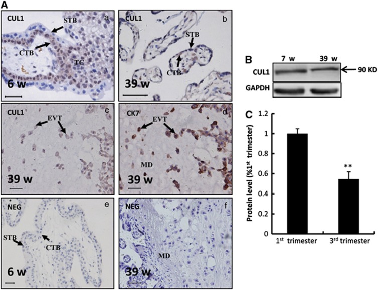
Expression of CUL1 in human placental villi at the first and the third trimesters. (A) Immunostaining of CUL1 and cytokeratin 7 (CK7) in normal human placental villi from the first and the third trimesters. (a) A placental villi from the first trimester. (b) A placental villi from the third trimester. (c, d) EVT invaded into the maternal decidua at the third trimester immunostained with CUL1 (c) and CK7, a marker of EVT (d). (e, f) Negative controls (NEG) in which normal IgG were used in place of primary antibody. CTB: cytotrophoblast cells; STB: syncytiotrophoblast; TC: trophoblastic column; EVT: extravillous trophoblast; MD: maternal decidua; Bar=100 μm. (B) Western blotting of CUL1 in the human placental villi from the first (n=3) and the third (n=3) trimesters. GAPDH was used as a loading control (here and after). (C) Three experiments as in (B) were quantified by measuring the intensity of CUL1 protein bands relative to GAPDH controls (t-test; **P<0.01)

