Figure 2.
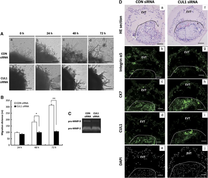
Silencing of CUL1 suppresses trophoblast outgrowth in extravillous explant cultures. (A) Extravillous explants from 5–8 weeks of gestation were maintained in culture on matrigel under low oxygen (3% O2) tension. Serial pictures of explants incubated with CUL1 siRNA or control (CON) siRNA were taken under the light microscope after 24, 48, and 72 h of culture in vitro. Bar=100 μm. (B) A statistical assay of the migration distance of villous tips. Data are presented as mean±S.D. of three independent experiments (t-test; *P<0.05; **P<0.01). (C) Serum-free culture medium of the explant transfected with indicated siRNAs was collected for gelatin zymography assay. (D) Examination of CUL1 knockdown efficiency after siRNA transfection. (a, f) Hematoxylin and eosin (HE) staining of CON or CUL1 siRNA-treated explants. Bar=100 μm. (b, g) Immunofluorescent staining using integrin α5 antibody (green) identifies the EVT. Bar=50 μm (c, h) Immunofluorescent staining using CK7 antibody (green) identifies the cytotrophoblast and EVT. Bar=50 μm (d, i) Immunofluorescent staining using CUL1 antibody (green) shows that CUL1 protein is lower in CUL1 siRNA-treated EVT (i) as compared with CON siRNA treated ones (d) at the same exposure time. Bar=50 μm. (e, j) DAPI staining of sections d and i to visualize nuclei. Bar=50 μm. a, b, c, and d are serial sections, so are f, g, h, and i. The region of EVT outgrowths is above the dashed lines

