Figure 6.
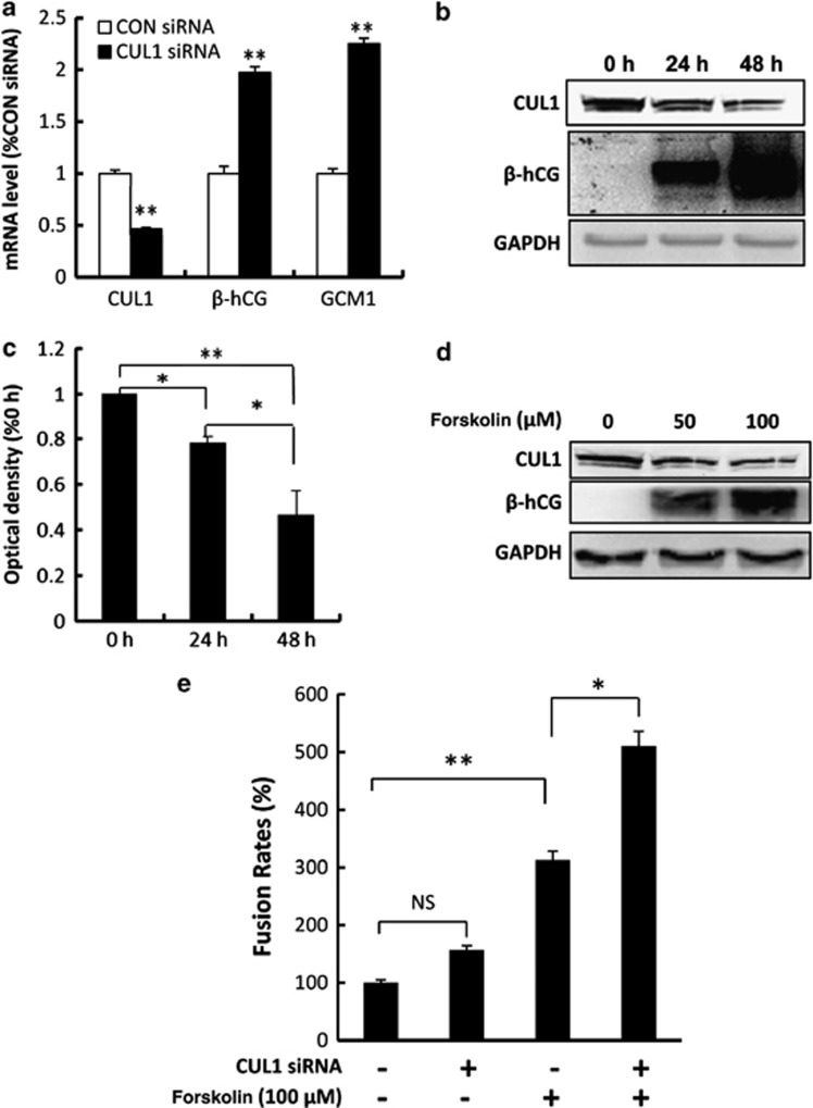
CUL1 protein level is decreased during syncytialization. (a) Real-time quantitative PCR analysis of CUL1, β-hCG, and GCM1 in the primary cultured cells treated with indicated siRNAs. The transcript level is expressed as percentage of CON siRNA, after normalization with β-actin (t-test; **P<0.01). (b) Western blotting showing the expression of CUL1 and β-hCG in the primary trophoblast cells cultured for 0 h to 48 h. (c) Statistical analysis of the western blotting results in (b) (one-way ANOVA; *P<0.05; **P<0.01). (d) Western blotting showing the expression of CUL1 in BeWo cells treated with or without forskolin by using indicated antibodies. (e) Twenty-four hours after transfection with siRNAs (CON and CUL1), BeWo cells were treated with 100 μM forskolin or methanol for 48 h. Multinucleated cells were counted as indicated in ‘Materials and Methods'. Statistical bar graph showed that CUL1 enhanced forskolin-induced syncytialization of BeWo cells (NS, P>0.05; *P<0.05; **P<0.01; ANOVA)

