Figure 2.
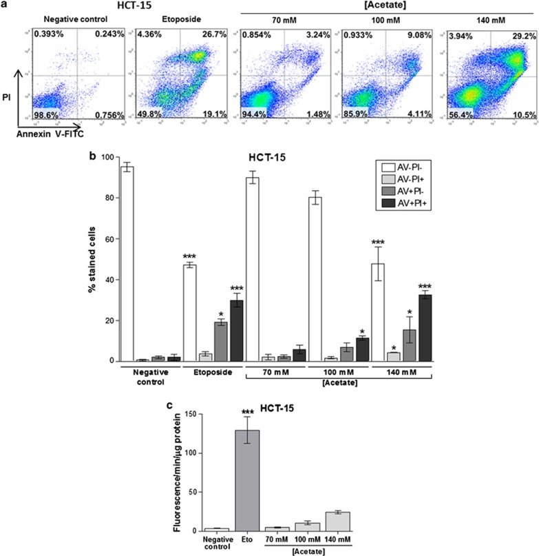
Acetate induces apoptosis and not necrosis in CRC cells. Apoptosis determined by Annexin V fluorescein isothiocyanate (AV-FITC) and propidium iodide (PI) assay in HCT-15 cells after incubation with 70 mM, 100 mM and 140 mM of acetate for 48 h. Cells were incubated with fresh complete medium or etoposide (50 μM) as a negative and positive control, respectively. (a) Representative histograms of HCT-15 cells double-labeled with AV and PI. Percentages of apoptotic cells (positive for AV) are the sum of the lower and upper right panels. (b) Quantitative analysis of AV/PI staining in HCT-15 cells. Values represent mean±S.E.M. of at least three independent experiments. *P≤0.05; ***P≤0.001, comparing each subset of cells (AV−PI−, AV−PI+, AV+PI−, AV+PI+) to the respective negative control cells. (c) Quantitative analysis of caspase 3 activity in HCT-15 cells after incubation with 70 mM, 100 mM and 140 mM of acetate for 48 h. Values represent mean±S.E.M. of three independent experiments. *P≤0.05; **P≤0.01; ***P≤0.001, compared with negative control cells

