Figure 6.
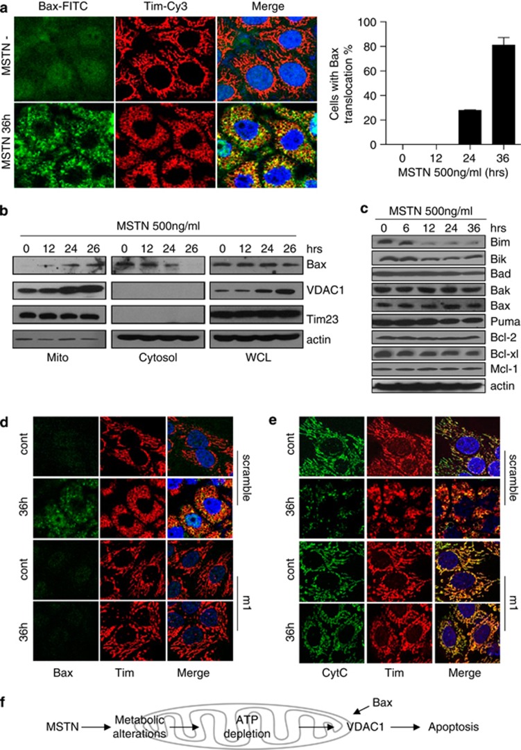
Mitochondrial translocation of Bax is involved in VDAC1-mediated apoptosis, which is induced by myostatin. (a) Left panel: HeLa cells treated with myostatin for 36 h were fixed and stained with anti-BAX (green) and anti-TIM (red) antibodies. Right panel: Statistics of HeLa cells with redistributed Bax in the presence of myostatin for 36 h. The data were shown as mean±S.E.M. (b) Subcellular localization of VDAC1 and Bax were examined by western blotting. Tim was shown as a loading control of mitochondrial sample; actin was shown as an loading control of cytosolic fraction and whole cell lysate. (c) Expression profile of major members of Bcl-2 family. (d) Scramble and siVD-m1 cells were treated with PBS or myostatin for 36 h. Immunofluorescence was performed with antibodies for Bax (green) and Tim (red). (e) HeLa cells were treated as in (d). Green: Cytochrome C; Red: Tim-Cy3. (f) A model for the mechanism underlying myostatin induction of VDAC1-mediated apoptosis in cancer cells

