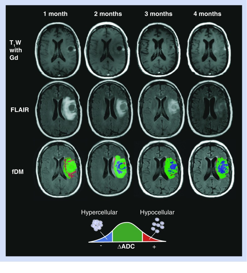
Figure 6. Standard MRI and functional diffusion maps in a patient with progressive disease after treatment with bevacizumab.
A 47-year-old male with a history of glioblastoma multiforme completed radiotherapy with concurrent temozolomide, followed by adjuvant temozolomide. His tumor recurred radiographically just prior to baseline ADC maps. The patient was then changed to bevacizumab monotherapy, and initially contrast enhancement and FLAIR signal abnormality improved substantially. The patient declined neurologically over 4 months of bevacizumab treatment, despite a positive radiographic response on postcontrast T1W (top row) and FLAIR images (middle row). The patient expired 2 months from the last fDM time point (6 months after start of bevacizumab treatment). During bevacizumab treatment, fDMs showed a rapid increase in the volume of hypercellularity (blue voxels), indicative of failed treatment.
ADC: Apparent diffusion coefficient; fDM: Functional diffusion map; FLAIR: Fluid-attenuated inversion recovery; T1W: T1 weighted.
Reproduced with permission from Springer (License No 2898871275280) [43].

