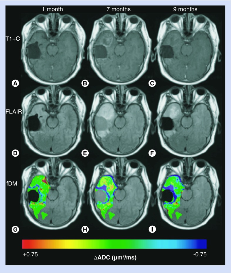
Figure 7. Changes in apparent diffusion coefficient predict progression in patient with suspected pseudoprogession.
Shown are postcontrast T1-weighted images (A–C), FLAIR images (D–F) and ADC changes (G–I) for a 52-year-old male patient diagnosed with a glioblastoma at 1 month (column 1), 7 months (column 2) and 9 months (column 3) post radiation therapy plus temozolomide. Based on standard imaging, pseudoprogression and radiation effect were presumed since enhancement appeared and then disappeared. Although true progression was not officially ruled out, radiology reports and discussions leaned more heavily towards a likely diagnosis of pseudoprogression and radiation effect. However, the ADC was progressively decreasing following radiotherapy, suggesting an increase in tumor cellularity, and thus true progression. The clinical course was consistent with true progression since the subject expired months following the scan shown in column 3.
ADC: Apparent diffusion coefficient; fDM: Functional diffusion map; FLAIR: Fluid-attenuated inversion recovery.

