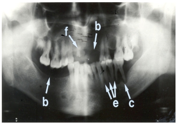
Fig. 3.
Panoramic radiograph showing more abnormalities than Fig. 2. This radiograph shows edentulous areas (i.e. loss of teeth) involving the right mandibular molar region and the left maxillary incisor and canine regions (b). A retained root of the right maxillary canine tooth is also present (f). Interdental bone loss is seen between the left mandibular premolar and molar teeth (e). A peri-apical radiolucency is associated with the left first mandibular molar tooth (c). The overall condition of the teeth of this patient is poor.

