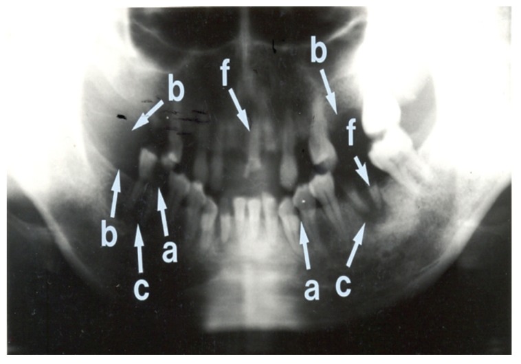
Fig. 4.
Panoramic radiograph showing many abnormalities. This radiograph shows multiple retained roots (f) and missing teeth (b) in both mandibular and maxillary arches. The right mandibular first molar tooth shows a large carious lesion with loss of crown enamel (a). The left mandibular canine teeth and the left mandibular first premolar tooth show caries interproximally (a). Peri-apical radiolucencies are noted in relation to the right and left mandibular molar teeth (c). The overall condition of the teeth of this patient is extremely poor.

