Figure 6.
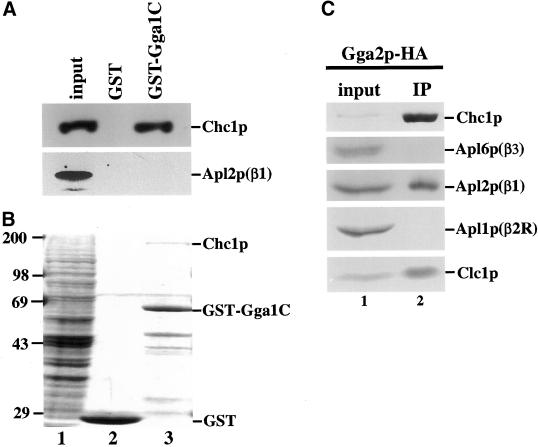
Interaction of Gga proteins and clathrin in vitro. The C-terminal half of Gga1p (aa 282–585) fused to GST (GST-Gga1C) or GST was bound to glutathione-Sepharose beads and incubated with extract from TVY614. Proteins bound to GST beads (lane 2) or GST-Gga1C beads (lane 3) were analyzed by SDS-PAGE followed by immunoblotting (A) or Coomassie blue staining (B) and compared with extract from TVY614 (input, lane 1). Inputs correspond to 100% (A) or 10% (B) of the amount of extract incubated with beads. (C) Extract from cells expressing Gga2p-3HA (GPY2373; lane 1) was subjected to nondenaturing immunoprecipitation with the use of anti-HA antibodies (IP, lane 2), followed by SDS-PAGE and immunoblotting. The input corresponds to 2% of the amount of extract subjected to immunoprecipitation.

