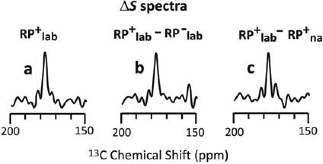
Figure 2.
13CO ΔS spectra based on the S0 and S1 spectra of three different ICP samples. The RP+lab and RP−lab plasmids respectively had and lacked the Fgp41 insert. The lab and na expression media respectively contained 13CO, 15N-Leu and unlabeled Leu. Panel a ΔS (RP+lab=S0–S1 signal represents directly-bonded 13CO-15N spin pairs of the RP+lab sample. Panel b ΔS=ΔS(RP+lab)–ΔS(RP−lab) is from spin pairs of IB Fgp41. Panel c ΔS=ΔS(RP+lab)–ΔS(RP+na) is from lab spin pairs of the RP+lab sample. The similar peak intensities, shifts, and lineshapes of the three spectra support a dominant contribution to the ΔS(RP+lab) spectrum of 13CO signals of labeled Ls of the LL dipeptides of IB Fgp41.

