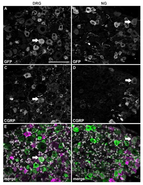
Figure 5.
Double immunolabeling for GFP and CGRP. Optical sections (epifluorescence and Apotome) of the DRG and NG double-labeled for GFP (A,B) and CGRP (C,D). DAPI staining appears in white in the merge color images (E,F). Note that double-labeled profiles (white arrows) are relatively rare in either ganglia. Scale bar = 100 μm (applies to all).

