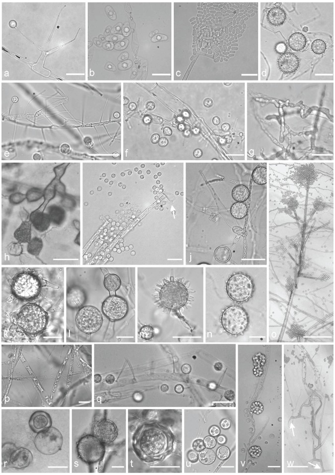
Fig. 2.
Typical morphological structures of different isolates of the Mortierellales, which are suitable for species delimitation. a. M. verticillata CBS 315.52, sporangiophore with a sporangiola; b. M. elongata FSU 9721, elongated sporangiospores containing central oil droplets; c. M. wolfii CBS 651.93, cracked sporangia releasing sporangiospores, on acrotonous branched tip of the sporangiophore; d. M. indohii CBS 720.71, stylospores; e. M. schmuckeri CBS 295.59, sporangiophores alongside a hypha with sporangiola; f. M. claussenii CBS 294.59, sporangiophores along a hypha with sporangiola; g. M. clonocystis CBS 357.76, typical swollen hyphae; h. M. zychae FSU 719, typical swollen hyphae arranged in clusters; i. M. parvispora FSU 10759, tip of a sporangiophore, sporangia leaving a collar (arrow), globose sporangiospores; j. M. lignicola CBS 207.37, sporangiophores, sporangiola (arrow 1), stylospores (arrow 2); k. M. exigua CBS 655.68, chlamydospores with typical outgrowing hyphae; l. M. gemmifera CBS 134.45, chlamydospores; m. M. hypsicladia CBS 116202, stylospores with projections; n. M. polygonia CBS 685.71, stylospores; o. M. nanthalensis CBS 610.70, acrotonous branching part of a sporangiophore; p. M. alpina FSU 2698, oil droplets containing hypha; q. M. camargensis CBS 221.58, sporangiophores along a hypha with sporangiola; r. M. epigama CBS 489.70, zygospores; s. M. echinosphaera CBS 575.75, chlamydospores; t. M. microszygospora CBS 880.97, microzygospore; u. M. camargensis CBS 221.58, oil droplets containing spheric sporangiola; v. Dissophora decumbens CBS 592.88, sporangiophores with sporangia; w. M. paraensis CBS 547.89, two sporangiophores with typical basitonous branchings (arrows mark the basal part). — Scale bars: a, b, i, n, p, r, u = 10 μm; c, j, q = 20 μm; d, e, g, h, m, v = 30 μm; f, k, l, s, t = 15 μm; o = 250 μm; w = 100 μm.

