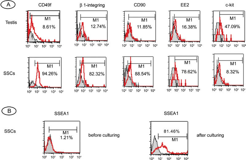
Figure 1.

Flow-cytometric characterization of human fetal testicular cells and human spermatogonial stem cells (SSCs). (A): Flow-cytometric characterization of human fetal testicular cells before and after magnetic cell sorting (MACS). The cells were stained with antibodies against CD49f, β1-integrin, CD90, EE2 and c-kit. The histograms are overlaid on relative isotype-matched control antibody histograms. Non-specific isotype staining controls are shown as shaded histograms; positively stained cells are shown as open histograms in red. Horizontal bars indicate mean percentages of positive cells from three independent experiments. (B): Flow-cytometric analysis of SSEA-1 expression in human SSCs before and after culture.
