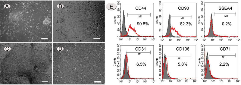
Figure 2.

Growth and characterization of hdFs. (A) – (D): Recovery of human embryonic stem cell-derived fibroblast-like cells (hdFs). (A): Morphology of human embryonic stem (hES) cell colonies after 4 days of co-culture with an MEF feeder layer. (B) and (C): Morphology of human embryonic stem cells (hESCs) maintained on matrigel without feeder layer support. (D): Morphology of isolated hdFs. Images were captured with an inverted Leica microscope using a 5 × objective with numerical aperture 0.12 and acquired through Spot camera and Spot software. (E): Flow-cytometric analysis of hdFs (passage 3) for the presence of CD44, CD90, SSEA-4, CD31, CD106 and CD71. The red line seems to be the positive signals from the antibodies and gray is the non-specific controls. Horizontal bars indicate mean percentages of positive cells from three independent experiments. (A) and (D): bars = 25 μm; (B) and (C), bars = 50 μm.
