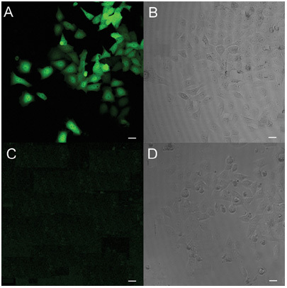
Figure 1.

Immunofluorescence staining of HEK293 cells transfected with pcDNA-hRNase9. The matched fluorescent (A, C) and phase-contrast images (B, D) of transfected HEK293 cells immunostained with anti-His antibody (A, B) and normal serum (C, D) are shown. Original magnification × 200. Bars = 20 μm.
