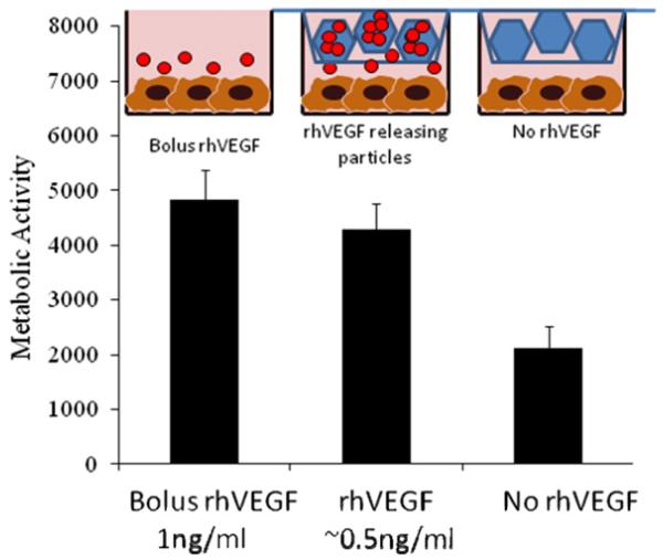
Fig. 7.
The release of rhVEGF from mineral-coated β-TCP granules significantly enhanced HUVEC proliferation, here measured using a blue cell titer assay (arbitrary units). There were no significant differences between bolus administration of rhVEGF and released rhVEGF, but, based on the release data, the concentration of rhVEGF from the slow release group was 0.5 ng ml−1. In the group exposed to bolus rhVEGF the cells were exposed to 1 ng ml−1. The granules releasing rhVEGF influenced proliferation at half the concentration of the bolus group.

