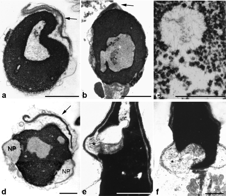
Figure 3.
Chromatin and acrosomal anomalies. The six panels depict spermatozoa with abnormal morphology and serious defects of chromatin condensation (nuclear lacunae). Nuclei show big areas of rarefaction with uncondensed chromatin of granular and fibrillar substructure (c). Some denser spots probably represent more condensed chromatin. Nuclear pockets (NP) are prominent (d) and show continuity with uncondensed chromatin (asterisks, e and f). Panels a and b show small acrosomes (acrosomal hypoplasia, arrows). In panels a and d small acrosomes are detached from nuclei (a and d). Bars=1 µm. b is reproduced with permission from Chemes.12

